
By Mark McConville
A SERIES of striking microscopic pictures has revealed the colourful patterns that make up the everyday illnesses the public is often struck down by.
Steve Gschmeissner / SPL / mediadrumimages.com
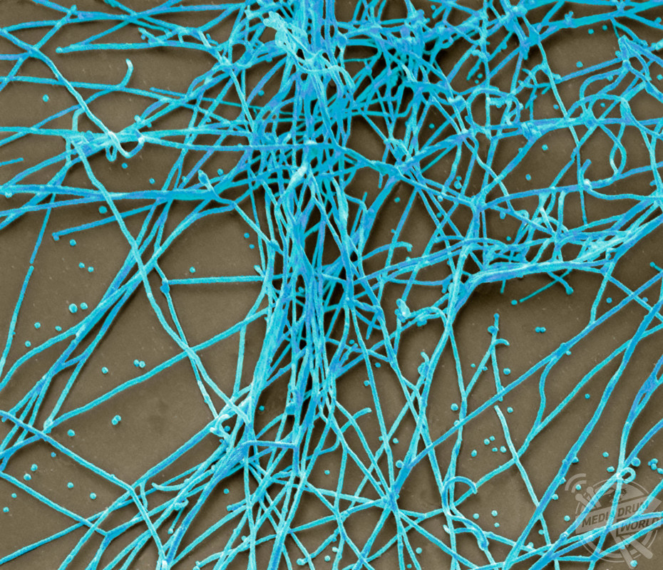
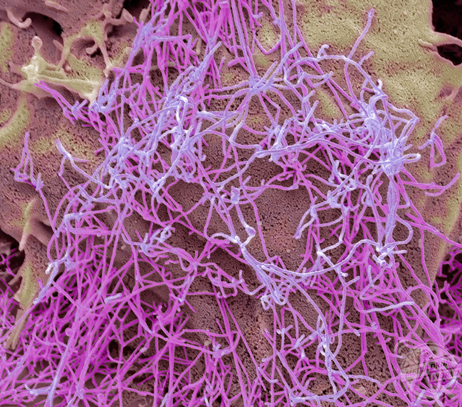
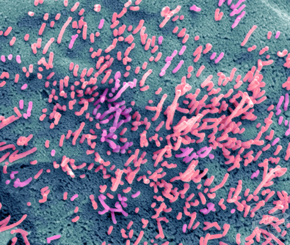
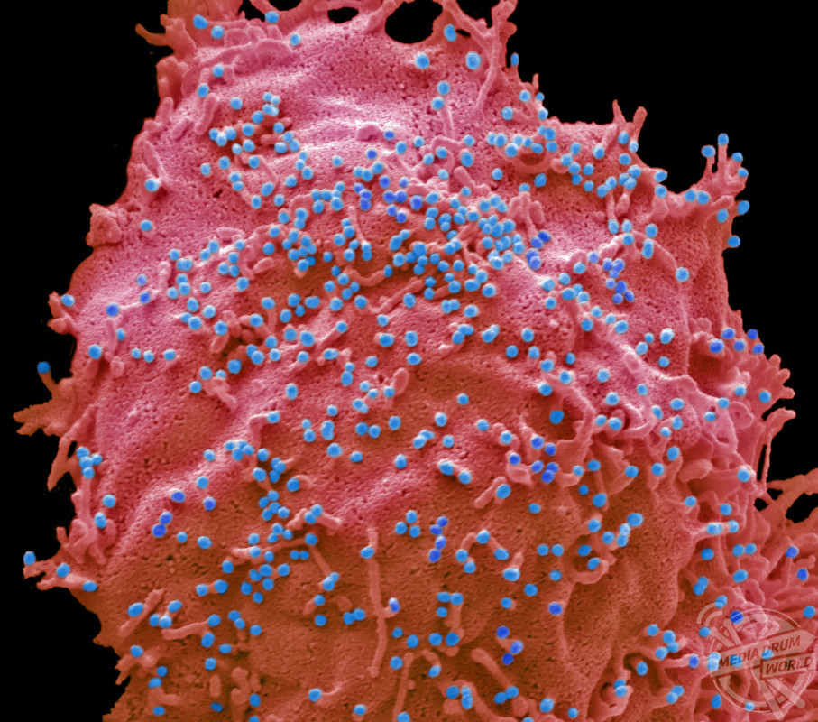
The incredible images show Aussie flu infection which looks like purple silly string, human cells infected with influenza B virus which looks like tiny seeds attaching themselves to a sponge and H1N1 flu strain infecting human cells.
Steve Gschmeissner / SPL / mediadrumimages.com
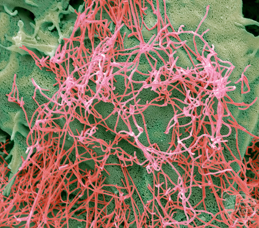
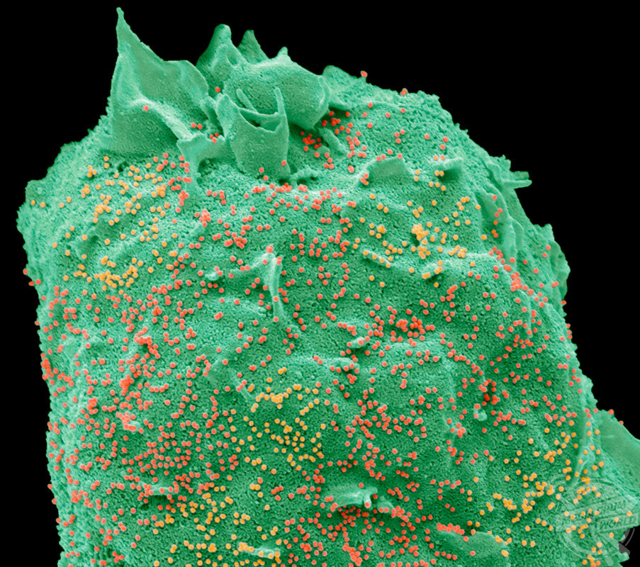
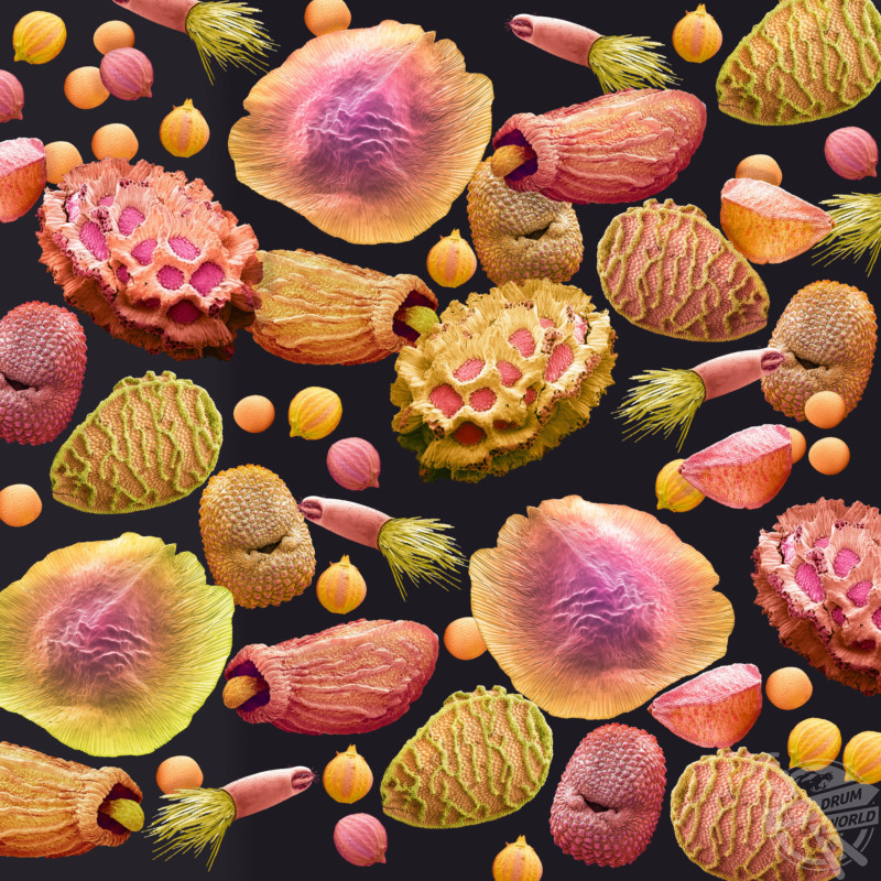
Other colourful snaps show the yellow and red capsules of the bird flu virus attacking human cells, seeds causing hayfever and the string-like threads of the H1N1 flu strain.
Steve Gschmeissner / SPL / mediadrumimages.com
The coloured scanning electron micrographs (SEM) were taken by Steve Gschmeissner who is one of the leading scanning electron microscopists in the world today.
Steve Gschmeissner / SPL / mediadrumimages.com
“For many years I have been waiting to share my wonderment at the microscopic world that exists around us and even in us,” he said.
Steve Gschmeissner / SPL / mediadrumimages.com
“For anyone involved in microscopy the SEM is the ultimate boy’s toy. Costing between £100,000 and £500,000 there are only a handful of people around the world who have access to this for fun. To be able to use this equipment is a dream come true.
Steve Gschmeissner / SPL / mediadrumimages.com
“The SEM picks up basically where the normal light microscope finishes. And it takes it so much further by magnifying the specimen up to a million times.
Steve Gschmeissner / SPL / mediadrumimages.com
“Also different to a regular microscope is the fact the SEM builds a 3D image using electrons giving you a unique view.”


