HomePopularAre You Brave Enough to Face These Parasites That Could be Hiding...
Are You Brave Enough to Face These Parasites That Could be Hiding in Your Home?
3083
Coloured scanning electron micrograph (SEM) of Brown recluse spider cephalothorax (Loxosceles reclusa). Characteristic features are the six eyes arranged in three pairs at the front of the head and the fiddle-shaped marking on the back. The brown recluse spider is often called the violin spider or fiddleback spider. The brown recluse bears a potentially deadly hemotoxic venom. Most bites are minor with no necrosis. However, a small number of brown recluse bites do produce severe dermonecrotic lesions. The bite of this spider is nasty and results in open, ulcerating sores. Left untreated such bites often become infected and significant tissue necrosis can occur. Magnification: x7 when shortest axis printed at 25 millimetres. The scans were taken by scientists Steve Gschmeissner, who is one of the world’s leading scanning electron microscopists in the world and award winning photo-micrographer Dennis Kunkel. SPL / mediadrumworld.com
Home Parasites
By Rebecca Drew
THESE frightening mini beasts that lurk in sofas and carpets on a mission to eat human skin will inspire you to give your home a good clean this winter.
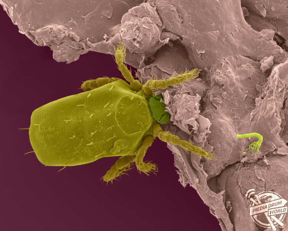
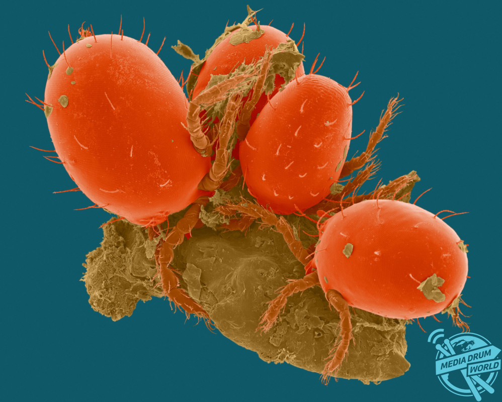
Coloured scanning electron micrograph (SEM) of Chigger, harvest mite larval ectoparasite (Trombicula sp.) on human epidermis (skin). Trombicula is a genus of harvest mites (also known as red bugs or berry bugs) from the Trombiculidae family. In their larval stage they are known as chiggers (or chigoe) and they attach to various animals, including humans, rabbits, toads, box turtles, quail, and even some insects. After crawling onto their hosts, they inject digestive enzymes into the skin that break down skin cells. They do not actually bite, but instead form a hole in the skin called a stylostome, and chew up tiny parts of the inner skin, thus causing severe irritation and swelling. This feeding process on skin causes severe itching. The larval stage is parasitic on humans and causes the disease called chigger dermatitis. Magnification: x34 when shortest axis printed at 25 millimetres. SPL / mediadrumworld.com
Stomach-churning images scrutinising the nasty parasites that could be lurking around your home have been revealed by a British boffin who collected samples from his own back garden.
The brightly coloured series of images show enlarged dust mites which can be found hiding on furniture and in carpets and chigger mites that can be found in fields and parks tucking all into human skin.
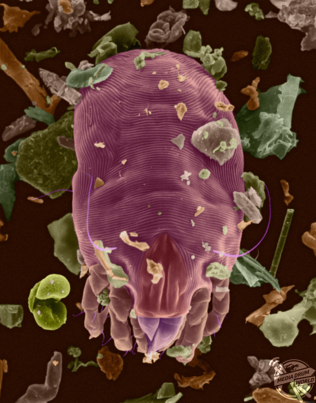
Coloured scanning electron micrograph (SEM) of Dust mite (Dermatophagoides sp.). Millions of dust mites inhabit the home, feeding on dead human skin that are common in house dust. The mite’s body is in three parts: the gnathosoma (head region) adapted for feeding on dead skin, the propodosma (carrying the 1st and 2nd pair of walking legs) and the hysterosoma (locating the 3rd and 4th pairs of legs). Dust mites produce 10-20 waste particles per day. The dead bodies and faecal pellets can trigger allergic responses. The whole life cycle from egg to adult takes approximately one month to complete, mature female mites can lay from 1-2 eggs per day. Adult mites can live up to two months. The most important house dust mites worldwide are Dermatophagoides farinae and Dermatophagoides pteronyssinus. Magnification: x91 when shortest axis printed at 25 millimetres. SPL / mediadrumworld.com
Other coloured scanning electron micrographs (SEM) show how these nasties could infiltrate our homes through our beloved pets. A dog tapeworm has been closely inspected as well as American dog ticks and a rabbit ear mite.
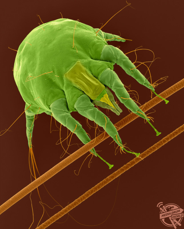
Coloured scanning electron micrograph (SEM) of Rabbit ear mite (Psoroptes cuniculi). Psoroptes cuniculi mites are non-burrowing, and chew/pierce the skin in the ear canal of rabbits causing psoroptic mange. Often a secondary bacteria infection develops, which can extend to the middle and inner ear causing torticollis. In addition, with a severe infestation, the mites may extend to the head, neck, and other parts of the body. These mites cause intense itching and often rabbits will scratch and shake their heads, which can lead to further infection. Their life span is approximately 21 days. Magnification: x35 when shortest axis printed at 25 millimetres. SPL / mediadrumworld.com
Another image shows what could be hanging out in our ponds and gardens, as a close-up of a bloodworm seemingly baring its teeth has also been captured.
In one picture, the bacteria on our seemingly clean chopping boards and kitchen sponges have also been magnified.
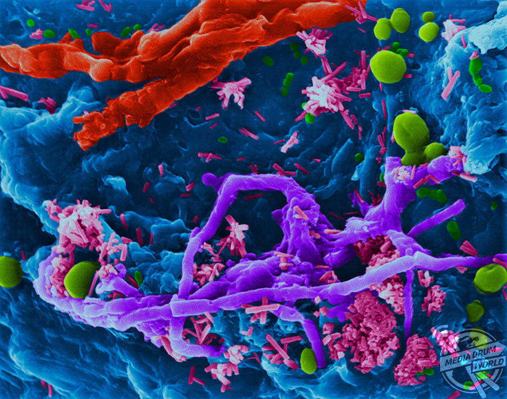
Coloured scanning electron micrograph (SEM) of Kitchen sponge microbes. Kitchen sponges can accumulate food and microbes when used for long periods of time and are not thoroughly cleaned. The moisture and food particles in a dirty sponge make it a perfect environment for microorganisms to grow (such as, bacteria and fungi). Their waste products give the sponge a distinctive smell. Features shown in this photomicrograph are: sponge surface (blue); bacteria (rod-shaped, purple and green colours); filamentous fungi (thin and thick filaments, purple and red colours); yeast fungi (round spheres, yellow, Green colour). Magnification: x580 when shortest axis printed at 25 millimetres. SPL / mediadrumworld.com
The scans were taken by scientists Steve Gschmeissner, who is one of the world’s leading scanning electron microscopists in the world and award winning photo-micrographer Dennis Kunkel.
“I basically look at anything that will fit in an SEM but parasites are especially fascinating and gruesome,” said Steve.
“I have looked at many hundreds of specimens and the bloodworms are from the mud in the bottom of my fishpond in the UK.”
Parasites are organisms that live in or on another living thing and feeds off of nutrients from its host.
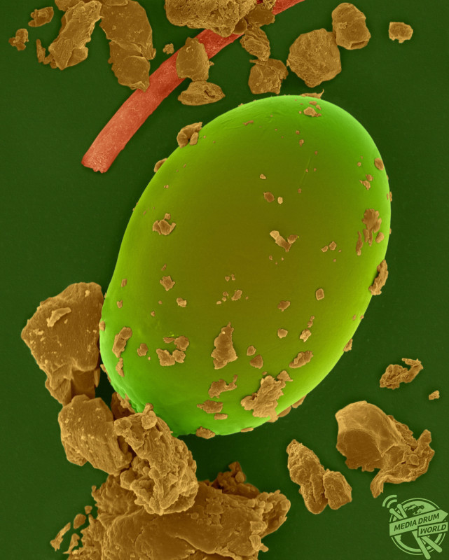
Coloured scanning electron micrograph (SEM) of Dust mite egg. (Dermatophagoides sp.). The whole life cycle from egg to adult takes approximately one month to complete, mature female mites can lay from 1-2 eggs per day. Adult mites can live up to two months. Millions of dust mites inhabit the home, feeding on dead human skin that are common in house dust. Dust mites produce 10-20 waste particles per day. The dead bodies and faecal pellets can trigger allergic responses. The whole life cycle from egg to adult takes approximately one month to complete, mature female mites can lay from 1-2 eggs per day. Adult mites can live up to two months. The most important house dust mites worldwide are Dermatophagoides farinae and Dermatophagoides pteronyssinus. Magnification: x200 when shortest axis printed at 25 millimetres. SPL / mediadrumworld.com
“Using electron microscopy methods and imaging reveals many things about bacterial morphology and human bacterial interactions,” said Dennis.
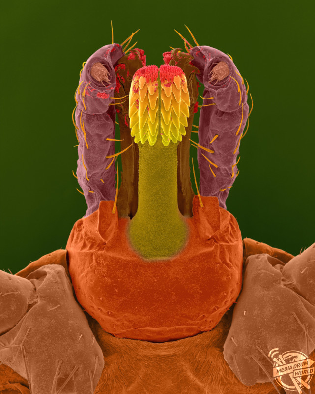
Coloured scanning electron micrograph (SEM) of Lone star tick head (Amblyomma americanum). A common ‘wood tick’ that includes other members, such as, the brown dog tick and the American dog tick. The mouthparts are a piercing and sucking type. The tick feeds by making a small incision in the skin with their barbed and piercing mouthparts. They insert their mouthparts and set the anchoring barbed teeth (hypostomer) in to the skin. They then secrete a fluid that cements their mouthparts into the skin. Lone star ticks live in wooded areas with underbrush, along creeks and rivers near animal resting places. Tick bites can be painful and cause localized inflammation, swelling and loss of blood. They may transmit disease agents, such as, ehrlichiosis, Rocky Mountain spotted fever, Lyme disease and tularemia. Magnification: x22 when shortest axis printed at 25 millimetres. SPL / mediadrumworld.com
It is widely thought that parasite infections are something that affect us after being on holiday but according to Cambridge Nutritional Scientists Ltd, ten percent of the UK’s population are contaminated by parasites at any one time.
Coloured scanning electron micrograph (SEM) of Chiggers (Trombicula sp.), harvest mite larval ectoparasite, on human epidermis (skin). Trombicula is a genus of harvest mites (also known as red bugs or berry bugs) from the Trombiculidae family. In their larval stage they are known as chiggers (or chigoe) and they attach to various animals, including humans, rabbits, toads, box turtles, quail, and even some insects. After crawling onto their hosts, they inject digestive enzymes into the skin that break down skin cells. They do not actually bite, but instead form a hole in the skin called a stylostome, and chew up tiny parts of the inner skin, thus causing severe irritation and swelling. This feeding process on skin causes severe itching. The larval stage is parasitic on humans and causes the disease called chigger dermatitis. Magnification: x26 when shortest axis printed at 25 millimetres. SPL / mediadrumworld.com